Yersiniosis in Fish – Yersinia ruckeri
Yersiniosis is a bacterial disease in fish caused by Yersinia ruckeri. While internationally, the disease is primarily problem in rainbow trout farming, outbreaks in Norway almost exclusively occur in farmed Atlantic salmon.

Pathogen and Transmission
Y. ruckeri occurs in multiple serotypes, but outbreaks in Norway are mainly linked to serotype O1, with occasional cases involving O2. Transmission is believed to take place primarily during the freshwater phase.
Outbreaks in the seawater phase typically occur shortly after transfer to sea, non-medicinal delousing, or other handling procedures, which may likely lead to stress-related activation of subclinical infections.
Research conducted by the Norwegian Veterinary Institute has identified a unique, genetic variant of Y. ruckeri serotype O1 as the cause of nearly all serious yersiniosis outbreaks in Norway since the mid-1990s. While this variant has not been documented outside of Norway, a different genetic variant has been identified as predominant in rainbow trout farming internationally. This variant has not yet been detected in Norway, although it was found in Swedish rainbow trout in 2017. Therefore, any detection of Y. ruckeri in Norwegian rainbow trout should be further investigated by the Norwegian Veterinary Institute to ensure early detection of a potential introduction.
Clinical Signs
Clinical signs of yersiniosis in farmed salmon are often non-specific and characteristic of septicaemia, including lethargy, respiratory distress, and abnormal swimming behaviour. Most outbreaks occur during the juvenile (freshwater) phase or shortly after sea transfer. Since 2016, however, outbreaks in the late seawater phase have become more common, especially in central Norway, prompting increased use of vaccination.
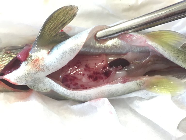
Other symptoms may include exophthalmia (bulging eyes), skin hyperpigmentation, ulcers, and haemorrhaging on the gills and at the base of the fins. Post-mortem findings may include abdominal fluid, small internal haemorrhages, splenomegaly (enlarged spleen), pale liver, and an empty gastrointestinal tract.
Diagnostics
Y. ruckeri can be easily cultured from the head kidney of affected fish and grows well at 20°C on standard media such as blood agar. Culture is recommended for reliable serotyping and genotyping. Histopathological findings are typically non-specific, but the bacterium can be confirmed through immunohistochemistry.
Occurrence
Since the first detection of Y. ruckeri in Norway in 1985, yersiniosis has been reported in Atlantic salmon farming operations along the entire Norwegian coastline.
Although a single genetic variant of Y. ruckeri serotype O1 is responsible for nearly all clinical cases in Norway, non-pathogenic (antivirulent) strains are widespread in freshwater hatcheries for Atlantic salmon. Thus, detection, e.g. in biofilm, of the bacterium in facilities without a history of disease does not necessarily indicate a clinical problem. Genotyping is necessary to determine whether an isolate belongs to the virulent variant.
Control Measures
Since the introduction of a commercial injectable vaccine against yersiniosis in 2020, its use has increased year by year. According to the Norwegian Veterinary Medicines Register (VetReg), approximately 230 million doses were delivered to smolt production facilities in 2023.
Antibiotic treatment may be used in some cases, though caution is advised due to the risk of developing antimicrobial resistance.
A product based on bacteriophages, i.e. viruses that specifically target Yersinia ruckeri, is available for control of the pathogen.
